|
All in all, I do realize that
you are more than likely concerned with appearance rather than function
although, with secondary and tertiary rhinoplasty loss or hindrance of
function is unfortunately a reality for many. From difficulty
breathing to headaches, from post-surgical nasal drip to depression -
there is more to rhinoplasty than aesthetics.
Understanding
The Structure Of The Nose
To understand how the appearance of the
nose is changed from within or to alleviate functionality problems, one
must understand how the external nose is supported and what must be done to the underlying nasal structure in
order to achieve these desires and goals. One must also realize that
changing one thing can, in turn, change another or at least affect it in
some way. This must always be taken into account so it is very important
to find a highly skilled surgeon to carry out your surgery. Revision
Rhinoplasty should be carried out to improve matters and not to worsen
them.
The
Nose According To A Genius - Henry Gray (1821–1865)
Henry Gray, a legend in his own time,
author of Anatomy of the Human Body - explains the human anatomy piece by
piece, cell by cell, function by function. It is Henry Gray that I believe
can best explain the structure of the nose with the aid of his
extraordinary engravings. I will include additional visual aids, in plain
English, to assist you in your understanding of the nose. *You will
more than likely need our Online
Rhinoplasty Glossary to translate the
terminology used. This section will load
in a new, smaller window for your convenience. You may want to keep it
open to ease of reference.
The Peripheral Olfactory Organ
"The peripheral olfactory organ
or organ of smell consists of two parts: an outer, the external
nose, which projects from the center of the face; and an internal,
the nasal cavity, which is divided by a septum into right
and left nasal chambers.
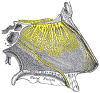
the External Nose (Nasus
Externus; Outer Nose)—The external nose is pyramidal in
form, and its upper angle or root is connected directly with the
forehead; its free angle is termed the apex. Its base is
perforated by two elliptical orifices, the nares [nostrils],
separated from each other by an antero-posterior septum, the columna [or
columella] . The margins of the nares are provided with a
number of stiff hairs, or vibrissæ, which arrest the passage of
foreign substances carried with the current of air intended for
respiration. The lateral surfaces of the nose form, by their union in
the middle line, the dorsum nasi [or simply,
dorsum], the direction of which varies considerably in
different individuals; the upper part of the dorsum is supported by the
nasal bones, and is named the bridge. The lateral surface ends
below in a rounded eminence, the ala nasi.
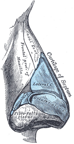
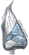
Structure.—The
frame-work of the external nose is composed of bones and cartilages; it
is covered by the integument, and lined by mucous membrane.
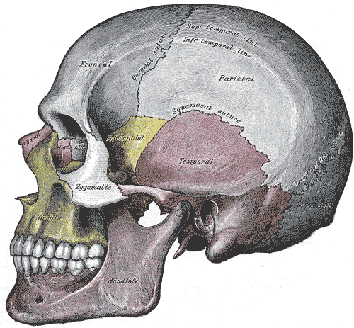
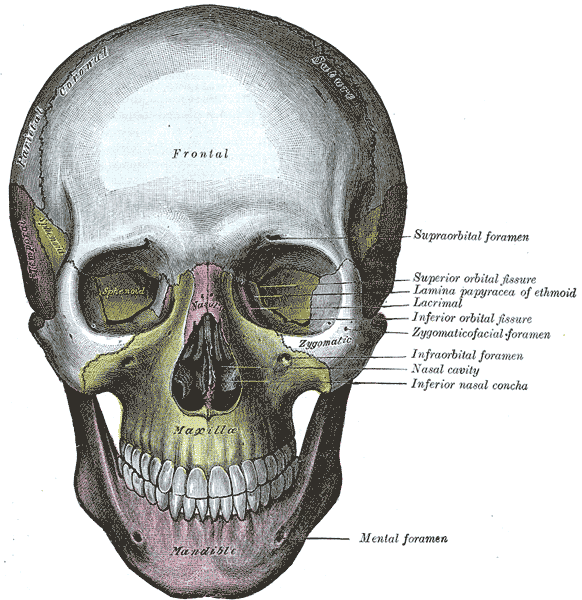
The bony frame-work
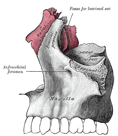
occupies the upper part of the organ; it consists of the nasal bones,
and the frontal processes of the maxillæ.
click diagrams for a
larger image
The cartilaginous
frame-work (cartilagines nasi) consists of five large pieces,
viz., the cartilage of the septum, the two lateral and the
two greater alar cartilages, and several smaller pieces, the lesser
alar cartilages. The various cartilages are connected to each
other and to the bones by a tough fibrous membrane.
cartilage
(lateral)
click diagram for a larger image
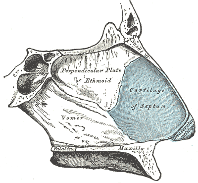
The cartilage of the
septum (cartilago septi nasi) is somewhat quadrilateral in
form, thicker at its margins than at its center, and completes the
separation between the nasal cavities in front. Its anterior margin,
thickest above, is connected with the nasal bones, and is continuous
with the anterior margins of the lateral cartilages; below, it is
connected to the medial crura of the greater alar cartilages by fibrous
tissue. Its posterior margin is connected with the perpendicular plate
of the ethmoid; its inferior margin with the vomer and the palatine
processes of the maxillæ.
 |
 |
| cartilage
(below) |
septum
(lateral) |
click diagrams for a
larger image
It may be prolonged backward
(especially in children) as a narrow process, the sphenoidal process,
for some distance between the vomer and perpendicular plate of the
ethmoid. The septal cartilage does not reach as far as the lowest part
of the nasal septum. This is formed by the medial crura of the greater
alar cartilages and by the skin; it is freely movable, and hence is
termed the septum mobile nasi.
The lateral cartilage
(cartilago nasi lateralis; upper lateral cartilage) is situated
below the inferior margin of the nasal bone, and is flattened, and
triangular in shape. Its anterior margin is thicker than the posterior,
and is continuous above with the cartilage of the septum, but separated
from it below by a narrow fissure; its superior margin is attached to
the nasal bone and the frontal process of the maxilla; its inferior
margin is connected by fibrous tissue with the greater alar cartilage.
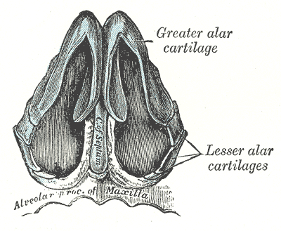
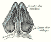
The greater alar
cartilage (cartilago alaris major; lower lateral cartilage)
is a thin, flexible plate, situated immediately below the preceding, and
bent upon itself in such a manner as to form the medial and lateral
walls of the naris of its own side. The portion which forms the medial
wall (crus mediale) is loosely connected with the
corresponding portion of the opposite cartilage, the two forming,
together with the thickened integument and subjacent tissue, the septum
mobile nasi. The part which forms the lateral wall (crus
laterale) is curved to correspond with the ala of the nose; it is
oval and flattened, narrow behind, where it is connected with the
frontal process of the maxilla by a tough fibrous membrane, in which are
found three or four small cartilaginous plates, the lesser alar
cartilages (cartilagines alares minores; sesamoid cartilages).
Above, it is connected by fibrous tissue to the lateral cartilage and
front part of the cartilage of the septum; below, it falls short of the
margin of the naris, the ala being completed by fatty and fibrous tissue
covered by skin. In front, the greater alar cartilages are separated by
a notch which corresponds with the apex of the nose.
The integument of the
dorsum and sides of the nose is thin, and loosely connected with the
subjacent parts; but over the tip and alæ it is thicker and more firmly
adherent, and is furnished with a large number of sebaceous follicles, the
orifices of which are usually very distinct
The arteries of the
external nose are the alar and septal branches of the external maxillary,
which supply the alæ and septum; the dorsum and sides being supplied from
the dorsal nasal branch of the ophthalmic and the infraorbital branch of
the internal maxillary. The veins end in the anterior facial and
ophthalmic veins.
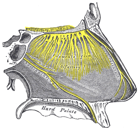
The nerves for the
muscles of the nose are derived from the facial, while the skin receives
branches from the infratrochlear and nasociliary branches of the
ophthalmic, and from the infraorbital of the maxillary.
nerve structure (lateral)
click diagram for a larger image
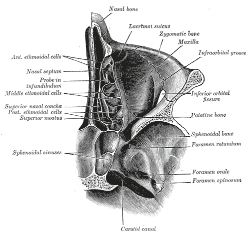
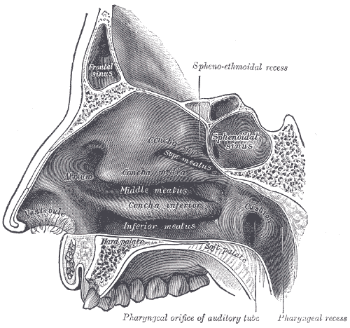
the Nasal Cavity (Cavum
Nasi; Nasal Fossa)—The nasal chambers are situated one on either
side of the median plane. They open in front through the nares, and
communicate behind through the choanæ with the nasal part of the pharynx.
The nares are somewhat pear-shaped apertures, each measuring about
2.5 cm. antero-posteriorly and 1.25 cm. transversely at its widest part.
The choanæ are two oval openings each measuring 2.5 cm. in the
vertical, and 1.25 cm. in the transverse direction in a well-developed
adult skull.
Inside the aperture of the
nostril is a slight dilatation, the vestibule, bounded laterally by
the ala and lateral crus of the greater alar cartilage, and medially by
the medial crus of the same cartilage. It is lined by skin containing
hairs and sebaceous glands, and extends as a small recess toward the apex
of the nose. Each nasal cavity, above and behind the vestibule, is divided
into two parts: an olfactory region, consisting of the superior
nasal concha and the opposed part of the septum, and a respiratory
region, which comprises the rest of the cavity."
Turbinate
Structures: More than Moisturizing
turbinate [2] (noun)
First appeared circa 1803
: one of usu. several thin plicated membrane-covered bony
or cartilaginous plates on the walls of the nasal chambers
This internal
structure is responsible for moisturizing the nose and the air that you
breathe. It can also change and control the temperature of the air that we
breathe into our sinus. If too cold and dry we can get headaches and
experience bleeding from dryness. If too damp from overproduction of
mucous we experience what is called rhinorrhea. I presently
have this and am probably going to have a portion of my turbinates
cauterized. I did not have this affliction until after my primary
rhinoplasty. Ever since my primary I have had a nonstop nasal drip. It is
said that I could be experiencing this from the primary infracture (or
bone breakage) to narrow the nose. It can also me due to scar tissue from
these infractures or from trauma.
 |
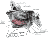 |
sinus
(lateral)
turbinates (conchae) |
Lateral
wall of right nasal cavity showing inferior concha in situ. |
*click diagrams for a
larger image*
Would
You Like To See Actual Photos Of The Turbinate Structures As Seen With
An Endoscope?
Another Excerpt from: Anatomy
of the Human Body by Henry Gray (1821–1865).
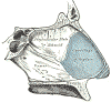
(Concha Nasalis Inferior;
Inferior Turbinated Bone)
The inferior nasal concha
extends horizontally along the lateral wall of the nasal cavity and
consists of a lamina of spongy bone, curled upon itself like a scroll.
It has two surfaces, two borders, and two extremities. The
medial surface is convex, perforated by numerous apertures, and
traversed by longitudinal grooves for the lodgement of vessels. The
lateral surface is concave,
and forms part of the inferior meatus. Its upper border is thin,
irregular, and connected to various bones along the lateral wall of the
nasal cavity. It may be divided into three portions: of these, the
anterior articulates with the conchal crest of the maxilla; the
posterior with the conchal crest of the palatine; the middle portion
presents three well-marked processes, which vary much in their size and
form. Of these, the anterior or lacrimal process is small and pointed
and is situated at the junction of the anterior fourth with the
posterior three-fourths of the bone: it articulates, by its apex, with
the descending process of the lacrimal bone, and, by its margins, with
the groove on the back of the frontal process of the maxilla, and thus
assists in forming the canal for the nasolacrimal duct. Behind this
process a broad, thin plate, the ethmoidal process, ascends to join the
uncinate process of the ethmoid; from its lower border a thin lamina,
the maxillary process, curves downward and lateralward; it articulates
with the maxilla and forms a part of the medial wall of the maxillary
sinus. The inferior border is free, thick, and cellular in structure,
more especially in the middle of the bone. Both extremities are more or
less pointed, the posterior being the more tapering.
References
Merriam-Webster Medical Dictionary
Graylab Online Medical Dictionary - UK
Anatomy of the Human Body, Henry Gray (1821–1865)
Online Version of Anatomy of the Human Body - Bartleby.com
Verbal and Online Interviews with:
-
Richard Ellenbogen, M.D.
-
James J. Romano, M.D.
University of Iowa Family
Practice Handbook, 3rd Edition, Chapter 19;
Otolaryngology: Nose; Mark A. Graber, M.D. and Laura Beaty, M.D.
Illustrated Encyclopedia of
Human Anatomic Variations:
-
Ronald A. Bergman, PhD
Department of Anatomy and Cell Biology
University of Iowa
Iowa City, Iowa, USA
- Adel K. Afifi, MD, MS
Departments of Pediatrics, Anatomy and Cell Biology, and Neurology
University of Iowa
Iowa City, Iowa, USA
- Ryosuke Miyauchi, MD
Department of Anatomy
Fukuoka University
Fukuoka, Japan
Olfactory Disorders, Dept. of
Otolaryngology, UTMB, Grand Rounds; November 17, 1993:
- Kelly Sweeny, M.D.
- Karen H. Calhoun, M.D.
- Melinda McCracken, M.S.
|